 Whiteboard Animation
Education · Biology lesson · 1:09
Animal cell animation
Watch an animal cell drawn step by step: the cell membrane, the nucleus in the middle, mitochondria, ribosomes, the Golgi body and small vacuoles. A hand-drawn whiteboard animation made from one illustration.
- Length
- 1:09 · 2 chapters
- Aspect ratio
- 4:3
- Colour starts at
- 0:55
- Style
- Lines + colour
- Formats
- 16:9 · 1:1 · 9:16
By Deniffer AlanPublished Tested by Deniffer Alan
Opens Studio with this picture, ready to draw.
Your own picture? How to prepare one
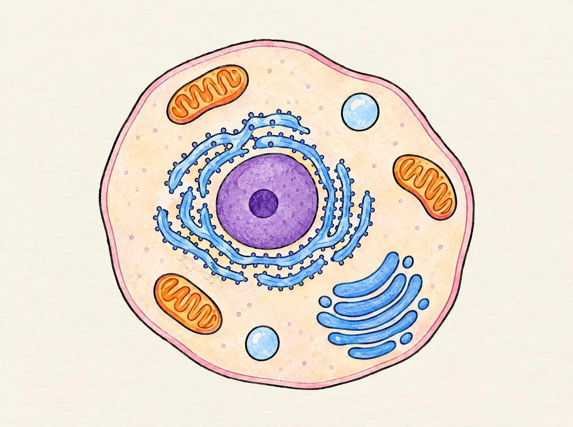
The picture it starts from
This finished illustration is all the drawing started from. Upload a picture like it and Studio draws it stroke by stroke; we don't generate pictures from text.
1920 × 1433 px · WEBP
The prompt behind the picture
Hand-drawn diagram of an animal cell cut open to show its inside: one large rounded, slightly irregular blob-shaped cell with a thin soft pink outer cell membrane and no cell wall. Inside, the pale cytoplasm holds: one large round purple nucleus near the middle with a darker nucleolus dot inside it, three bean-shaped orange mitochondria with folded wavy lines inside, a stack of curved flat blue sacs (Golgi apparatus), wavy folded ribbons of rough endoplasmic reticulum dotted with tiny ribosome dots wrapped beside the nucleus, and two small round vacuoles. No chloroplasts, no cell wall, no large central vacuole. Nothing else in the picture. Coloured pencil and watercolour marker, clean black ink outlines, warm off-white paper, no text.The picture came from an image model; any finished illustration works the same way, including one you drew. Use the prompt as a starting point for your own topic.
How this clip was made
The picture above was made with an image model from the prompt above; WhiteboardAnimation.ai then drew it, stroke by stroke, as the clip at the top of this page.
- Drawn
- Credits used
- 1
Notes from making it
Each part in the narration was checked against the Talking Glossary of the US National Human Genome Research Institute.
Animal cell, step by step
A script to read over it, timed to the drawing: the lines go down first and the colour arrives on the last beat.
- 0:00Line artYour body is built from tiny cells. Let’s look inside one animal cell.
- 0:11Around the outside is the cell membrane. It protects the cell and controls what goes in and out.
- 0:22Inside, a jelly-like liquid called cytoplasm fills the cell, and the other parts float in it.
- 0:33The big round nucleus sits in the middle. It holds the chromosomes, which carry the cell’s genetic instructions.
- 0:44ColourThe bean-shaped mitochondria make most of the energy the cell needs. Tiny dots called ribosomes build proteins, and the stacked Golgi body packs them up to send out.
- 0:56Animal cells have only small vacuoles, which help store waste. And unlike plant cells, they have no tough cell wall outside.
Where this animal cell animation fits
Use it in a biology lesson on cells, to start a topic comparing plant and animal cells, or as a short clip in a science video for kids. The same process turns your own diagram into a clip.
- Education
- Explainer videos
- Science
Questions