 Whiteboard Animation
Education · Biology lesson · 1:01
Respiratory system diagram animation
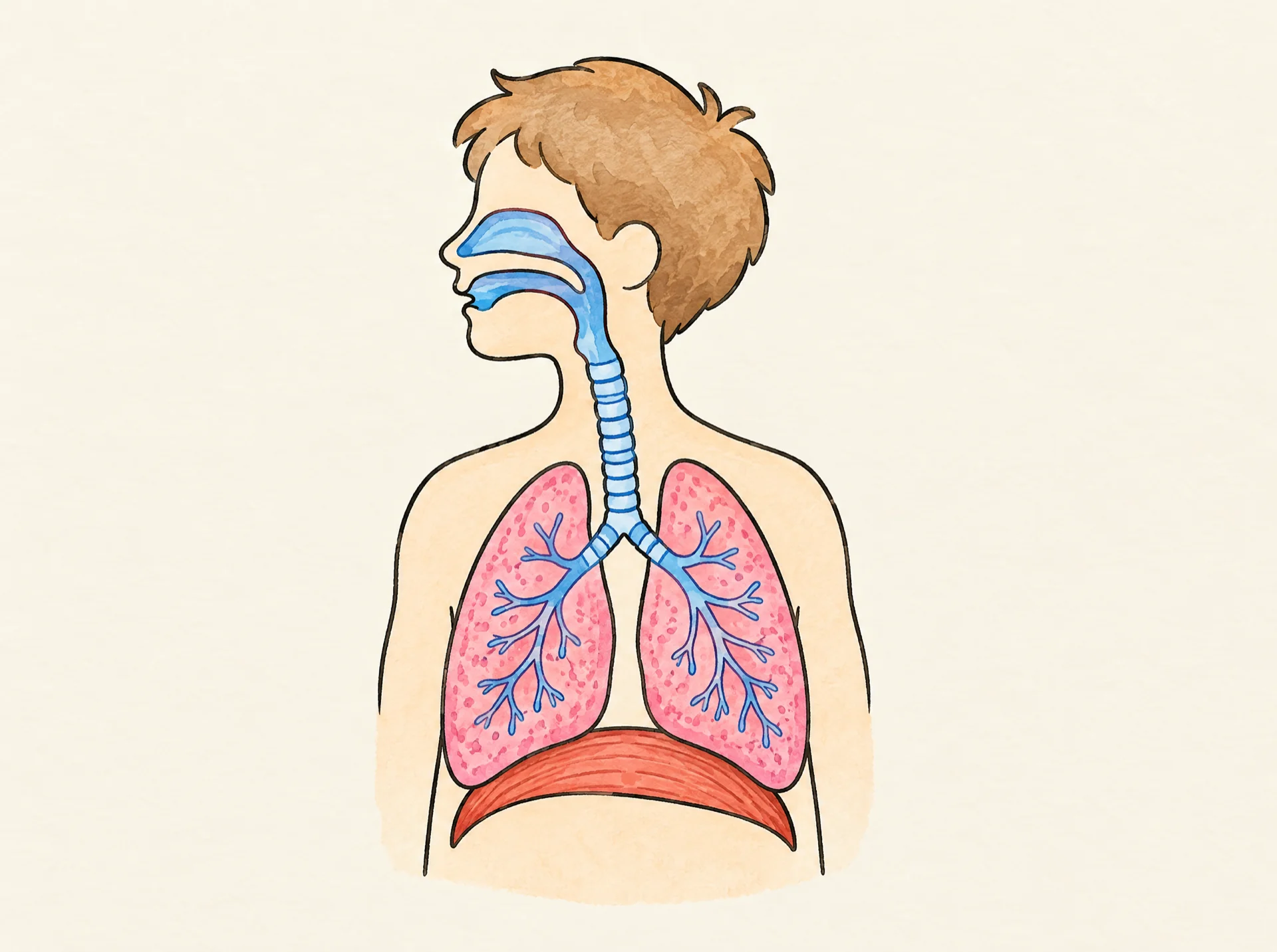
Watch a respiratory system diagram drawn step by step: the nose and mouth, the windpipe, two bronchi branching into the lungs and the dome-shaped diaphragm below. A hand-drawn whiteboard animation made from one illustration.
- Length
- 1:01 · 2 chapters
- Aspect ratio
- 4:3
- Colour starts at
- 0:26
- Style
- Lines + colour
- Formats
- 16:9 · 1:1 · 9:16
By Deniffer AlanPublished Tested by Deniffer Alan
Opens Studio with this picture, ready to draw.
Your own picture? How to prepare one
The picture it starts from
This finished illustration is all the drawing started from. Upload a picture like it and Studio draws it stroke by stroke; we don't generate pictures from text.
1920 × 1433 px · WEBP
The prompt behind the picture
Hand-drawn diagram of the human respiratory system inside a simple pale outline of a child's head, neck and chest, the head turned to the side as a cut-away so the air path shows: the nasal cavity behind the nose and the mouth both open into the throat. From the throat the windpipe (trachea) runs straight down the neck with ring-shaped ridges, then splits into exactly two bronchi, one going into each lung, branching into thin tubes inside. Two large pink spongy lungs. Under the lungs, one thin red muscle sheet, the diaphragm, shaped like a dome that bulges UPWARD into the chest like an upside-down bowl, and the bottoms of both lungs curve to rest on top of the dome. No heart, no ribs, no other organs. Nothing else in the picture, plenty of empty space around the body. Coloured pencil and watercolour marker, clean black ink outlines, warm off-white paper, no text.The picture came from an image model; any finished illustration works the same way, including one you drew. Use the prompt as a starting point for your own topic.
How this clip was made
The picture above was made with an image model from the prompt above; WhiteboardAnimation.ai then drew it, stroke by stroke, as the clip at the top of this page.
- Drawn
- Credits used
- 1
Notes from making it
The first picture drew the diaphragm curving down like a smile; it was made again with the dome bulging up under the lungs. The narration was checked against MedlinePlus from the US National Library of Medicine and the American Lung Association.
Respiratory system, step by step
A script to read over it, timed to the drawing: the lines go down first and the colour arrives on the last beat.
- 0:00Line artEvery cell in your body needs oxygen. Your respiratory system brings oxygen in and takes extra carbon dioxide away.
- 0:05Air comes in through your nose or mouth and moves into your throat.
- 0:11Then it travels down the windpipe, or trachea. This strong tube has rings that stop it from collapsing.
- 0:16The trachea splits into two bronchi, one for each lung. They branch into smaller and smaller tubes that end in tiny air sacs called alveoli.
- 0:21ColourIn the air sacs, oxygen moves into your blood, and carbon dioxide moves out to be breathed away.
- 0:27Under the lungs is the diaphragm, a dome-shaped muscle. When it tightens, air is pulled in through your nose or mouth.
Where this respiratory system diagram animation fits
Use it in a biology lesson on breathing, before measuring breaths after exercise, or as a short clip in a health video for kids. The same process turns your own diagram into a clip.
- Education
- Explainer videos
- Science
Questions